无机材料学报 ›› 2013, Vol. 28 ›› Issue (1): 109-116.DOI: 10.3724/SP.J.1077.2013.12109
马玉菲, 乔 莉, 冯庆玲
收稿日期:2012-03-23
修回日期:2012-05-29
出版日期:2013-01-10
网络出版日期:2012-12-20
作者简介:马玉菲(1987–), 女, 博士研究生. E-mail: mayf09@mails.tsinghua.edu.cn
基金资助:MA Yu-Fei, QIAO Li, FENG Qing-Ling
Received:2012-03-23
Revised:2012-05-29
Published:2013-01-10
Online:2012-12-20
About author:MA Yu-Fei. E-mail: mayf09@mails.tsinghua.edu.cn
Supported by:摘要:
碳酸钙广泛存在于生物矿物中, 是地球上最普遍的生物矿物之一。贝壳和珍珠的主要组成部分为碳酸钙无机相。我国淡水养殖珍珠多数品质优异, 具有良好的珍珠光泽。该种珍珠以文石晶型碳酸钙为无机相, 称为文石珍珠。近年来, 在我国淡水养殖珍珠中发现了球文石的存在, 球文石的出现是导致珍珠失去光泽、降低质量的主要原因。本文对比阐述了淡水文石珍珠和球文石珍珠的微观结构与性能, 总结了与珍珠层有关的体外模拟碳酸钙生物矿化的实验结果, 提出了珍珠层生物矿化机理未来的研究方向。
中图分类号:
马玉菲, 乔 莉, 冯庆玲. 淡水珍珠的生物矿化机理研究进展[J]. 无机材料学报, 2013, 28(1): 109-116.
MA Yu-Fei, QIAO Li, FENG Qing-Ling. Research Progress on Biomineralization Mechanism of Freshwater Pearl[J]. Journal of Inorganic Materials, 2013, 28(1): 109-116.
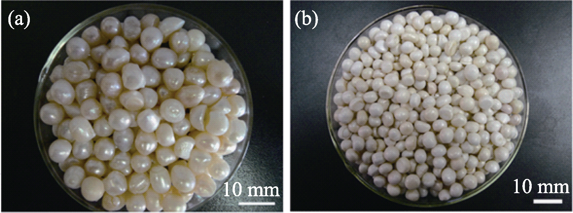
图1 文石珍珠(a)和球文石珍珠(b)的光学照片
Fig. 1 Optical photos of aragonite pearls (a) and vaterite pearls (b)
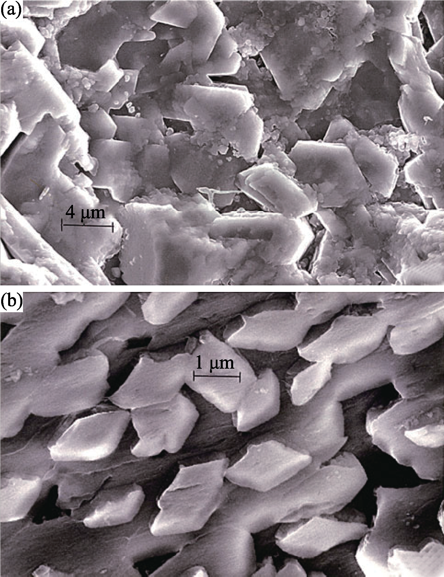
图2 珍珠内层晶体(方解石)(a)及外层晶体(文石)(b)的扫描电镜照片[19]
Fig. 2 Comparative SEM views for pearl internal layer crystals (mostly calcite) (a) and external surface crystals (mostly aragonite) (b)[19]
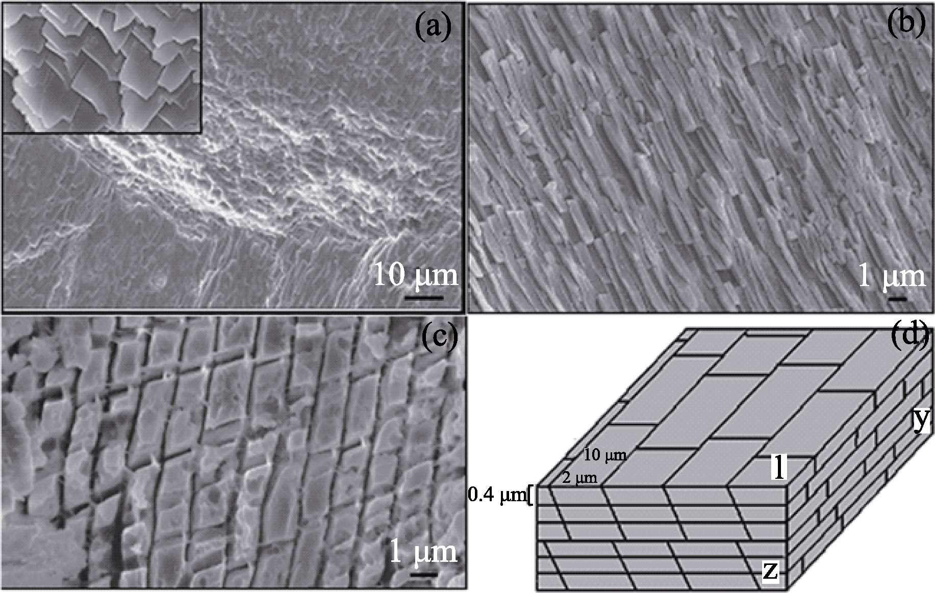
图3 球文石珍珠层的三维微观结构和示意图[20]
Fig. 3 SEM images of vaterite tablets and layers in lackluster pearls after 10wt% EDTA-2Na treatment[20]
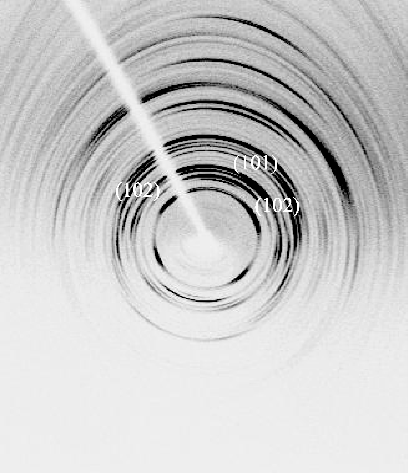
图4 球文石珍珠表面微区XRD分析[22]
Fig. 4 X-ray microanalysis of vaterite pearl surface[22]

图5 球文石板片内部的HRTEM形貌(a)和对应的SAED结果(b)[23]
Fig. 5 HRTEM micrograph (a) and SAED corresponding patterns (b) of the vaterite pearl. Curve A indicates twin stacking faults, and curve B stands for superstructure. The SAED patterns show the superstructure and disordered stacking[23]

图6 文石珍珠水可溶有机质(a)及球文石珍珠酸可溶有机质(b)诱导的碳酸钙晶体[37]
Fig. 6 Calcium carbonate crystals induced by WSM of aragonite pearl (a) and ASM of vaterite pearl (b)[37]
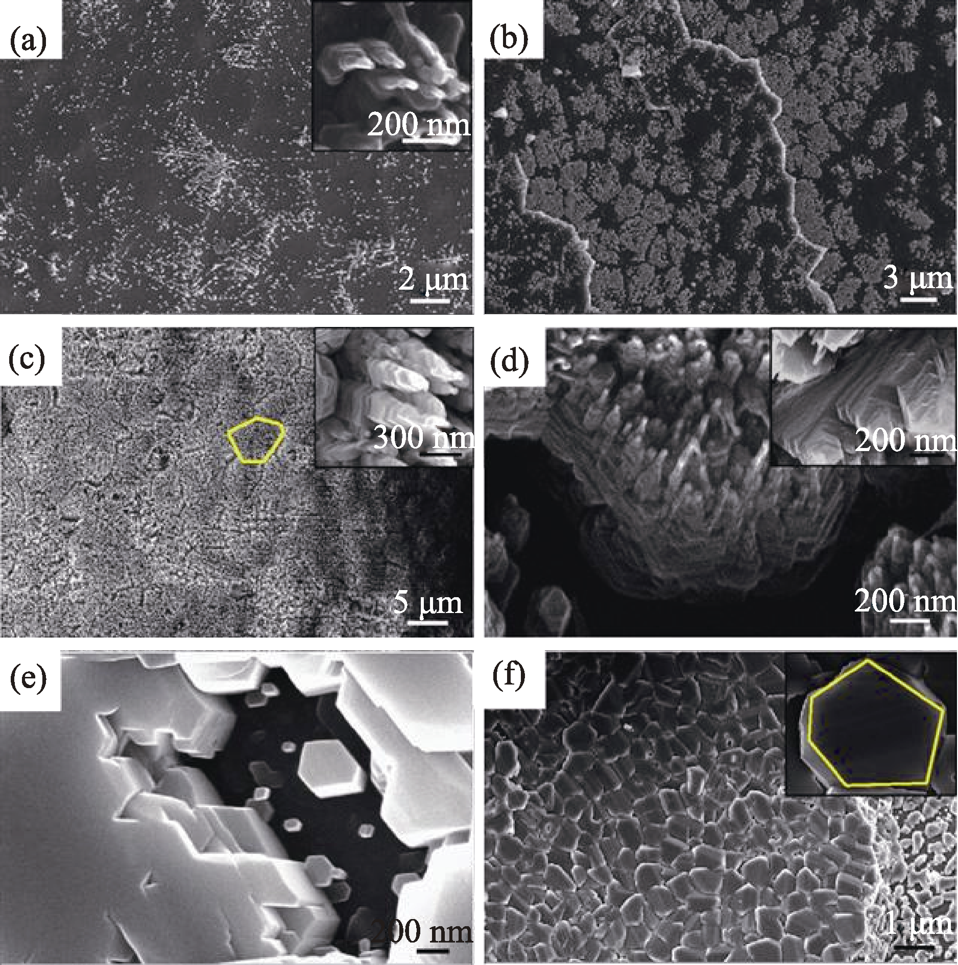
图7 “类珍珠层板片”的生长过程[42]
Fig. 7 SEM images of CaCO3 grown on nacre surface for different time[42]
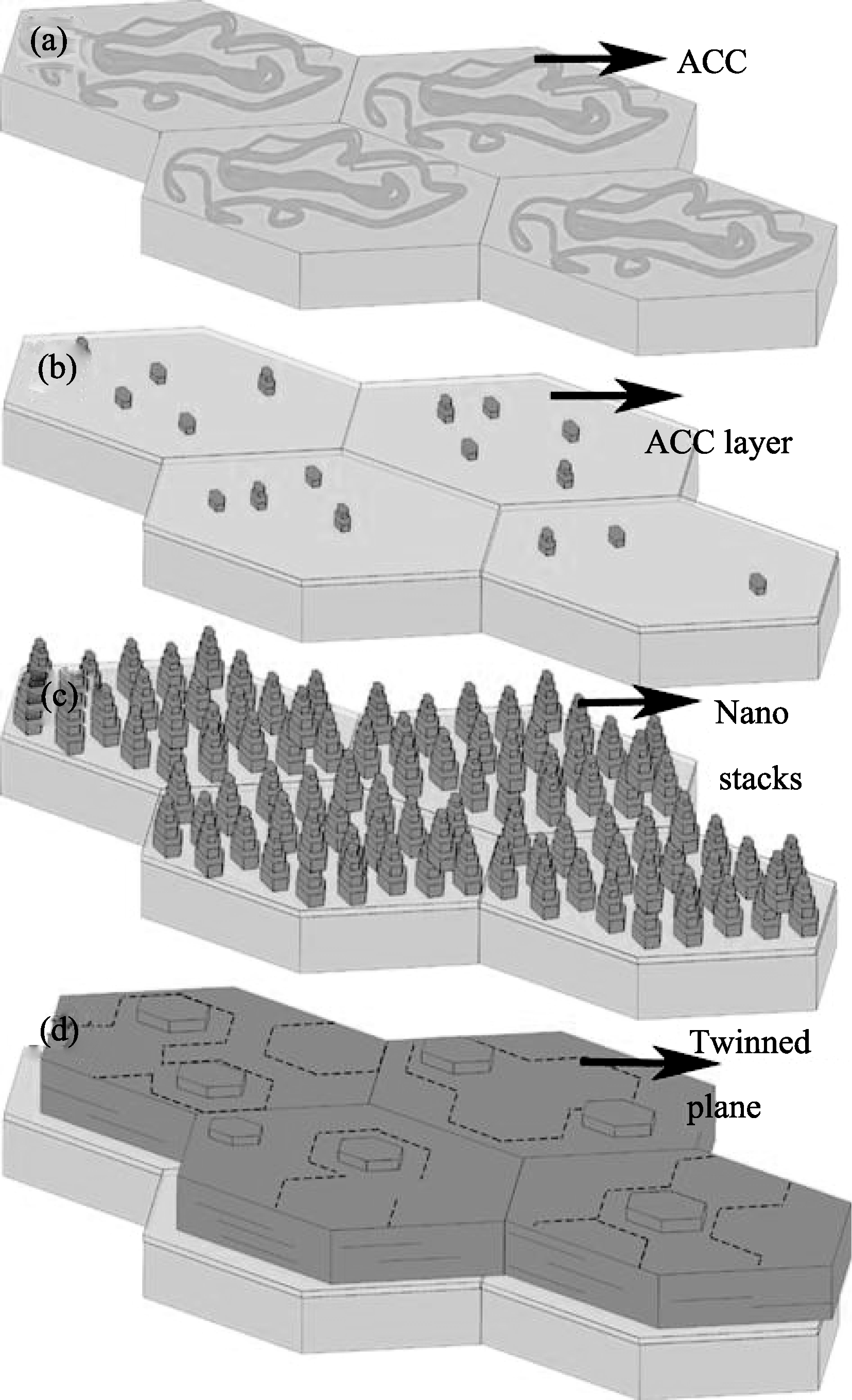
图8 原始珍珠层上碳酸钙晶体生长模型图[42]
Fig. 8 Schematic representation of the growth sequence of CaCO3 on nacre[42]
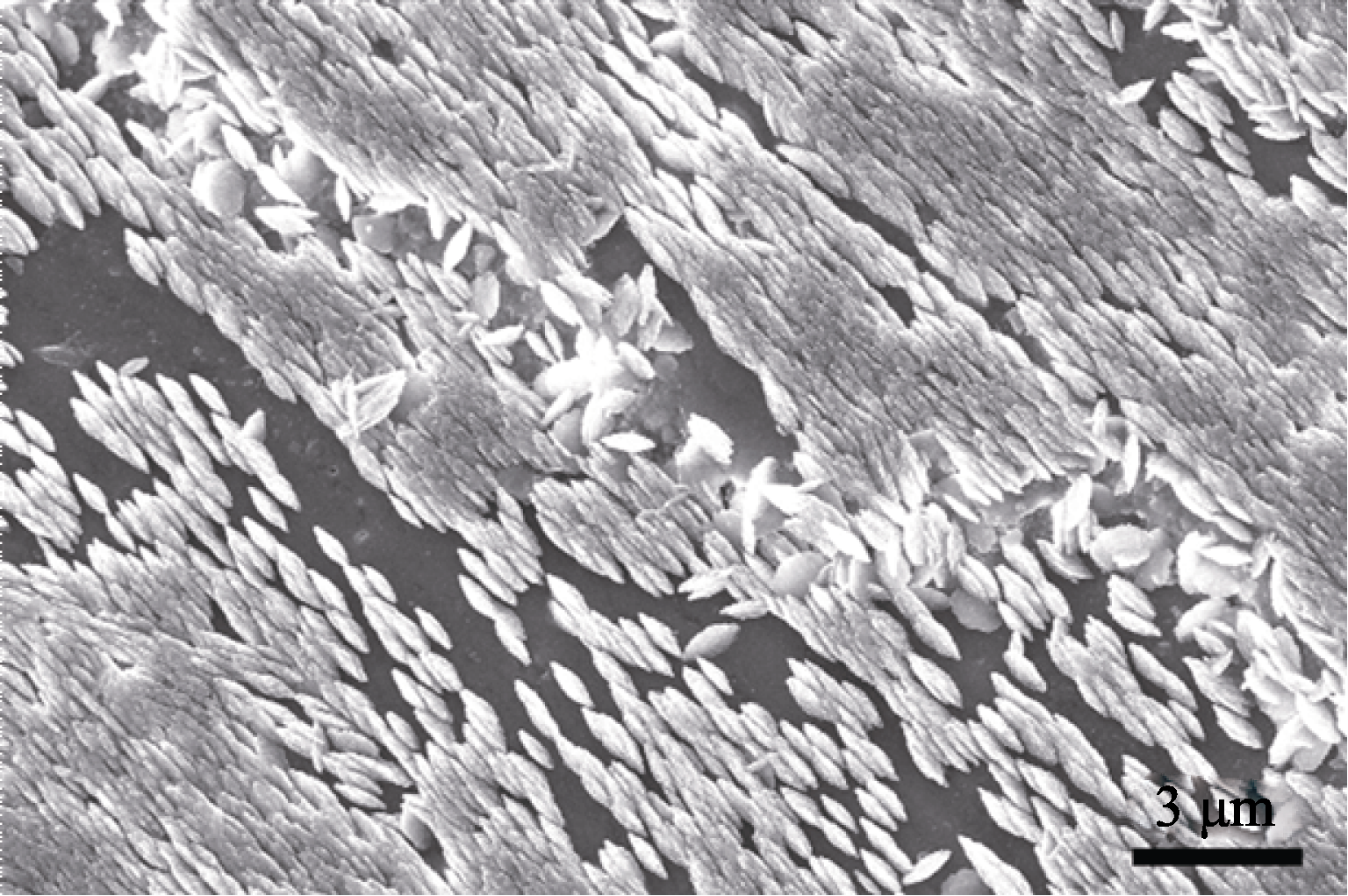
图9 在球文石珍珠层表面生长的球文石晶体的SEM照片[22]
Fig. 9 SEM image of vaterite crystals grown on the original section of vaterite pearl[22]
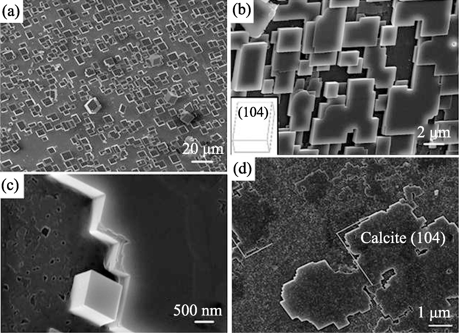
图10 谷氨酸改性的方解石基底上生长的方解石晶体[43]
Fig. 10 SEM micrographs of calcite matrix and calcite layers deposited on glutamic acid layers[43]
| [1] | 崔福斋. 生物矿化. 北京: 清华大学出版社, 2007: 13-14, 54-61, 330-336. |
| [2] | Rosenberg G D, Hughes W W, Parker D L. The geometry of bivalve shell chemistry and mantle metabolism. Am. Malacol. Bull., 2001, 16(1/2): 251-261. |
| [3] | Tang M, Shi A J. Overview of studies on calcium metabolism in molluscs. Journal of Fisheries of China, 2000, 24(1): 86-91. |
| [4] | Watabe N. Crystal growth of calcium carbonate in the invetebrate. Prog. Cryst. Growth Ch., 1981, 4: 99-147. |
| [5] | Nakahara H, Bevelander G. Electromicroscopic and amino acid studies on the outer and inner shell layers of Haliotis rufescena. Venus, 1982, 41(1): 33-46. |
| [6] | Nakahara H. An electromicroscope study of the growing surface of nacre in two gastropod spiecies Turbo cornutus and Tegula pfeifferi. Venus, 1979, 38(3): 205-211. |
| [7] | Weiner S, Hood L. Soluble protein of organic matrix of mollusk shells: a potential template for shell formation. Science, 1975, 190(5): 987-988. |
| [8] | Weiner S, Traub W. Macromolecules in mollusc shells and their function in biomineralization. Philos. Trans. R Soc. B-Biol. Sci., 1984, 304(1121): 425-433. |
| [9] | Addadi L, Joester D, Nudelman F, et al. Mollusk shell formation: a source of new concepts for understanding biomineralization processes. Chem-Eur. J, 2006, 12(4): 981-987. |
| [10] | Ma H Y, Dai T G, Yuan K R, et al. The first discovery of vaterite in lusterless fresh-water pearl of Leidian, Zhejiang. Acta Mineralogica Sinica, 2001, 21(2): 153-157. |
| [11] | Zhang G S, Xie X D. Utrastructure and formation theory of nacre shells. J. Mineral. Petrol., 2000, 2(1): 11-16. |
| [12] | Gutmannsbauer W, Hanni H A. Structural and chemical investigation on shells and pearls of nacre forming salt-and fresh-water bivalve molluscs. J. Gemm., 1994, 24(4): 241-252. |
| [13] | Ren F Z, Wan X D, Ma Z H, et al. Study on microstructure and thermodynamics of nacre in mussel shell. Mater. Chem. Phys., 2009, 114(1): 367-370. |
| [14] | Schaffer T E, Ionescu-Zanetti C, Proksch R, et al. Does abalone nacre form by heteroepitaxial nucleation or by growth through mineral bridges?Chem. Mater., 1997, 9(8): 1731-1740. |
| [15] | Zaremba C M, Belcher A M, Fritz M, et al. Critical transitions in the biofabrication of abalone shells and flat pearls. Chem. Mater., 1996, 8(3): 679-690. |
| [16] | Manne S, Zaremba C M, Giles R, et al. Atomic-force microscopy of the nacreous layer in mollusk shells. Prog. Cryst. Growth Ch., 1994, 256(1345): 17-23. |
| [17] | Checa A G, Rodriguez-Navarro A B. Self-organisation of nacre in the shells of Pterioida (Bivalvia : Mollusca). Biomaterials, 2005, 26(9): 1071-1079. |
| [18] | Feng Q L, Cui F Z, Li H D. Crystal orientation, toughening mechanisms and a mimic of nacre. Mat. Sci. Eng. C, 2000, 11(1): 19-25. |
| [19] | Murr L E, Ramirez D A. The microstructure of the cultured freshwater pearl. JOM, 2012, 64(4): 469-474. |
| [20] | Qiao L, Feng Q L, Li Z. Special vaterite found in freshwater lackluster pearls. Cryst. Growth & Des., 2007, 7(2): 275-279. |
| [21] | Zhang G S, Hao Y L. Microscopic morphology of porcelaneous layers on surface of freshwater cultured pearl. J. Gems Gemmol., 2004, 6(1): 1-3. |
| [22] | 乔 莉. 淡水球文石珍珠结构及其矿化机理研究. 北京: 清华大学博士论文, 2008. |
| [23] | Qiao L, Feng Q L. Study on twin stacking faults in vaterite tablets of freshwater lacklustre pearls. J. Cryst. Growth, 2007, 304(1): 253-256. |
| [24] | Grasby S E. Naturally precipitating vaterite (mu-CaCO3) spheres: Unusual carbonates formed in an extreme environment. Geochim. Cosmochim. Acta, 2003, 67(9): 1659-1666. |
| [25] | Qiao L, Feng Q L, Liu Y. A novel bio-vaterite in freshwater pearls with high thermal stability and low dissolubility. Mater. Lett., 2008, 62(12/13): 1793-1796. |
| [26] | Lakshminarayanan R, Chi-Jin E O, Loh X J, et al. Purification and characterization of a vaterite-inducing peptide, pelovaterin, from the eggshells of Pelodiscus sinensis (Chinese soft-shelled turtle). Biomacromolecules, 2005, 6(3): 1429-1437. |
| [27] | Takahashi K, Yamamoto H, Onoda A, et al. Highly oriented aragonite nanocrystal-biopolymer composites in an aragonite brick of the nacreous layer of Pinctada fucata. Chem. Comm., 2004(8): 996-997. |
| [28] | Tang R K, Orme C A, Nancollas G H. A new understanding of demineralization: the dynamics of brushite dissolution. J. Phys. Chem. B, 2003, 107(38): 10653-10657. |
| [29] | Levi-Kalisman Y, Falini G, Addadi L, et al. Structure of the nacreous organic matrix of a bivalve mollusk shell examined in the hydrated state using Cryo-TEM. J. Struct. Biol., 2001, 135(1): 8-17. |
| [30] | Crenshaw M A. The soluble matrix from Mercenaria mercenaria shell. Biomineralization, 1972, 6: 6-11. |
| [31] | Thompson J B, Paloczi G T, Kindt J H, et al. Direct observation of the transition from calcite to aragonite growth as induced by abalone shell proteins. Biophys. J., 2000, 79(6): 3307-3312. |
| [32] | Feng Q L, Pu G, Pei Y,et al. Polymorph and morphology of calcium carbonate crystals induced by proteins extracted from mollusk shell. J. Cryst. Growth, 2000, 216(1-4): 459-465. |
| [33] | Belcher A M, Wu X H, Christensen R J, et al. Control of crystal phase switching and orientation by soluble mollusc-shell proteins. Nature, 1996, 381(6577): 56-58. |
| [34] | Falini G, Albeck S, Weiner S, et al. Control of aragonite or calcite polymorphism by mollusk shell macromolecules. Science, 1996, 271(5245): 67-69. |
| [35] | Samata T, Hayashi N, Kono M, et al. A new matrix protein family related to the nacreous layer formation of Pinctada fucata. Febs. Lett., 1999, 462(1/2): 225-229. |
| [36] | Kono M, Hayashi N, Samata T. Molecular mechanism of the nacreous layer formation in Pinctada maxima. Biochem. Biophys. Res. Commun., 2000, 269(1): 213-218. |
| [37] | Ma Y F, Qiao L, Feng Q L. In-vitro study on calcium carbonate crystal growth mediated by organic matrix extracted from fresh water pearls. Mat. Sci. Eng. C, 2012, 32(7):1963-1970. |
| [38] | Ma Y F, Gao Y H, Feng Q L. Effects of pH and temperature on CaCO3 crystallization in aqueous solution with water soluble matrix of pearls. J. Cryst. Growth, 2010, 312(21): 3165-3170. |
| [39] | Hou W T, Feng Q L. Morphologies and growth model of biomimetic fabricated calcite crystals using amino acids and insoluble matrix membranes of Mytilus edulis. Cryst. Growth Des., 2006, 6(5): 1086-1090. |
| [40] | Nassif N, Pinna N, Gehrke N, et al. Amorphous layer around aragonite platelets in nacre. Proc. Natl. Acad. Sci., 2005, 102(36): 12653-12655. |
| [41] | Rousseau M, Lopez E, Stempflé P, et al. Multiscale structure of sheet nacre. Biomaterials, 2005, 26(31): 6254-6262. |
| [42] | Qiao L, Feng Q L, Lu S S. In vitro growth of nacre-like tablet forming: from amorphous calcium carbonate, nanostacks to hexagonal tablets. Cryst. Growth & Des., 2008, 8(5): 1509-1514. |
| [43] | Qiao L, Feng Q L, Lu S S. Alternate deposition of oriented calcite and amino acid layer on calcite substrates. J. Phys. Chem. B, 2008, 112(43): 13635-13640. |
| [1] | 丁玲, 蒋瑞, 唐子龙, 杨运琼. MXene材料的纳米工程及其作为超级电容器电极材料的研究进展[J]. 无机材料学报, 2023, 38(6): 619-633. |
| [2] | 杨卓, 卢勇, 赵庆, 陈军. X射线衍射Rietveld精修及其在锂离子电池正极材料中的应用[J]. 无机材料学报, 2023, 38(6): 589-605. |
| [3] | 陈强, 白书欣, 叶益聪. 热管理用高导热碳化硅陶瓷基复合材料研究进展[J]. 无机材料学报, 2023, 38(6): 634-646. |
| [4] | 林俊良, 王占杰. 铁电超晶格的研究进展[J]. 无机材料学报, 2023, 38(6): 606-618. |
| [5] | 牛嘉雪, 孙思, 柳鹏飞, 张晓东, 穆晓宇. 铜基纳米酶的特性及其生物医学应用[J]. 无机材料学报, 2023, 38(5): 489-502. |
| [6] | 苑景坤, 熊书锋, 陈张伟. 聚合物前驱体转化陶瓷增材制造技术研究趋势与挑战[J]. 无机材料学报, 2023, 38(5): 477-488. |
| [7] | 杜剑宇, 葛琛. 光电人工突触研究进展[J]. 无机材料学报, 2023, 38(4): 378-386. |
| [8] | 杨洋, 崔航源, 祝影, 万昌锦, 万青. 柔性神经形态晶体管研究进展[J]. 无机材料学报, 2023, 38(4): 367-377. |
| [9] | 游钧淇, 李策, 杨栋梁, 孙林锋. 氧化物双介质层忆阻器的设计及应用[J]. 无机材料学报, 2023, 38(4): 387-398. |
| [10] | 张超逸, 唐慧丽, 李宪珂, 王庆国, 罗平, 吴锋, 张晨波, 薛艳艳, 徐军, 韩建峰, 逯占文. 新型GaN与ZnO衬底ScAlMgO4晶体的研究进展[J]. 无机材料学报, 2023, 38(3): 228-242. |
| [11] | 陈昆峰, 胡乾宇, 刘锋, 薛冬峰. 多尺度晶体材料的原位表征技术与计算模拟研究进展[J]. 无机材料学报, 2023, 38(3): 256-269. |
| [12] | 齐占国, 刘磊, 王守志, 王国栋, 俞娇仙, 王忠新, 段秀兰, 徐现刚, 张雷. GaN单晶的HVPE生长与掺杂进展[J]. 无机材料学报, 2023, 38(3): 243-255. |
| [13] | 林思琪, 李艾燃, 付晨光, 李荣斌, 金敏. Zintl相Mg3X2(X=Sb, Bi)基晶体生长及热电性能研究进展[J]. 无机材料学报, 2023, 38(3): 270-279. |
| [14] | 刘岩, 张珂颖, 李天宇, 周菠, 刘学建, 黄政仁. 陶瓷材料电场辅助连接技术研究现状及发展趋势[J]. 无机材料学报, 2023, 38(2): 113-124. |
| [15] | 谢兵, 蔡金峡, 王铜铜, 刘智勇, 姜胜林, 张海波. 高储能密度聚合物基多层复合电介质的研究进展[J]. 无机材料学报, 2023, 38(2): 137-147. |
| 阅读次数 | ||||||
|
全文 |
|
|||||
|
摘要 |
|
|||||