Journal of Inorganic Materials ›› 2020, Vol. 35 ›› Issue (8): 867-881.DOI: 10.15541/jim20190561
Special Issue: 封面文章; 生物材料论文精选(2020)
• REVIEW • Previous Articles Next Articles
DONG Shaojie1,2( ),WANG Xudong2,SHEN Steve Guofang2,3,WANG Xiaohong1(),LIN Kaili2()
Received:2019-11-04
Revised:2019-11-24
Published:2020-08-20
Online:2020-01-20
Supported by:CLC Number:
DONG Shaojie,WANG Xudong,SHEN Steve Guofang,WANG Xiaohong,LIN Kaili. Research Progress on Functional Modifications and Applications of Bioceramic Scaffolds[J]. Journal of Inorganic Materials, 2020, 35(8): 867-881.
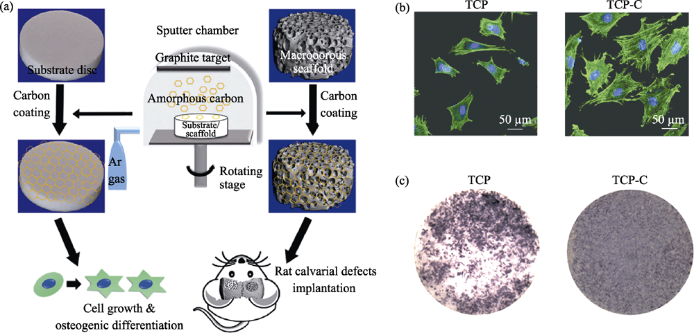
Fig. 1 Procedure for the fabricating of β-TCP scaffold coated with amorphous carbon (a), and adhesion (b) and ALP activity (c) of BMSCs cultured on the samples[18]
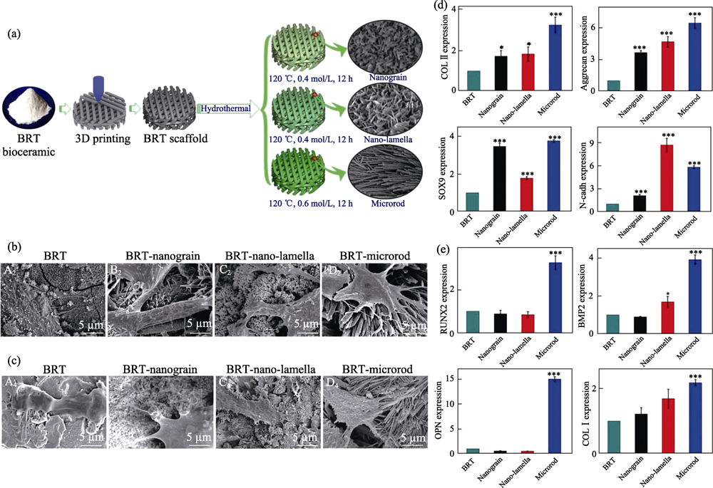
Fig. 2 (a) Fabrication procedure of bredigite (BRT) scaffolds with modified micro/nanostructure on the surface, cell adhesion behavior of (b) chondrocytes and (c) BMSCs cultured on different scaffolds, and expression level of (d) chondrogenesis of chondrocytes and (e) osteogenesis related genes of BMSCs cultured on different scaffolds, respectively[28]
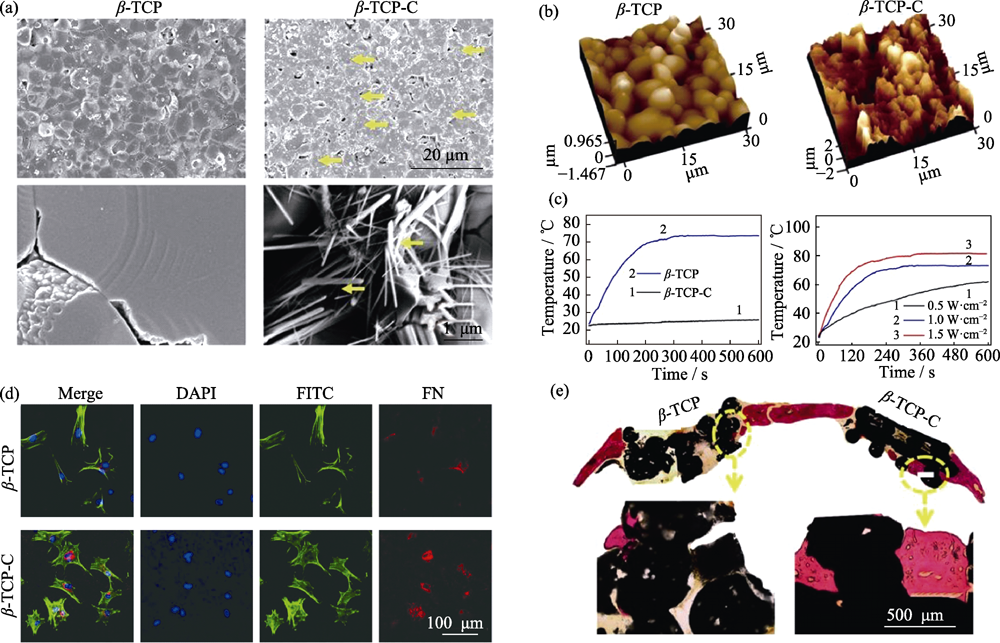
Fig. 3 Surface morphology of β-TCP and CA coated β-TCP (β-TCP-C) detected with (a) SEM (yellow, CA) and (b) atomic force microscope, (c) photothermal performance of β-TCP and β-TCP-C, (d) cell adhesion behavior and FN-expression of BMSCs cultured on β-TCP and β-TCP-C, and (e) osteogenesis capability of β-TCP and β-TCP-C scaffold in vivo[32]
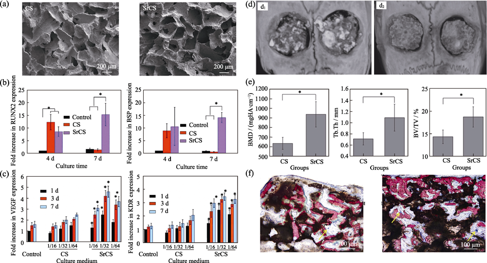
Fig. 4 (a) Morphologies of CS and Sr-CS scaffolds, expression level of (b) osteogenic genes of BMSCs-OVX and (c) angiogenic genes of HUVECs cultured with the extracts of CS and Sr-CS scaffolds, (d) micro-CT images (d1, CS; d2, Sr-CS), (e) morphometric analysis and (f) VG staining results of the new formed bone (left: CS; right: Sr-CS)[39]
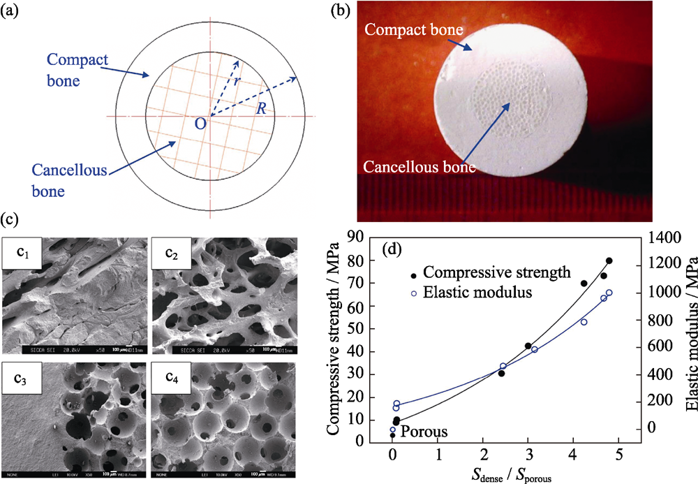
Fig. 5 (a) Structure-diagram and (b) digital images of biomimetic β-TCP scaffolds (c) SEM images of (c1-c2) compact/cancellous interface of natural bone and (c3-c4) dense/porous interface of scaffold, (d) function curves of compression strength, modulus of elasticity and dense/porous cross-sectional area ratio[61]
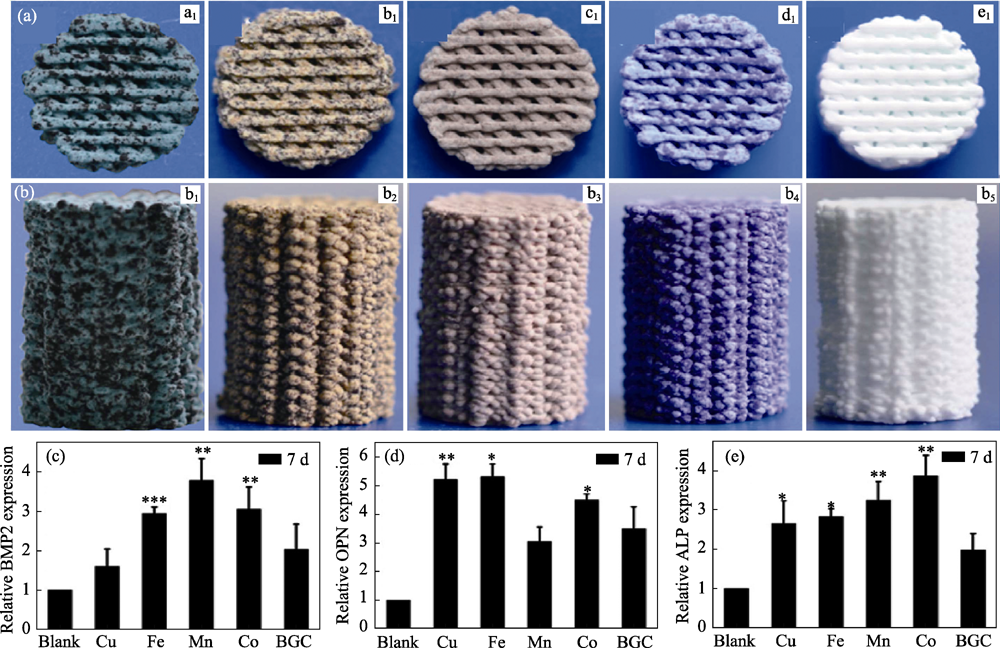
Fig. 6 Morphologies (top view (a) and side view (b)) of 3D printing bioglass scaffolds containing Cu, Fe, Mn, Co elements and pure bioglass, (c-e) expression of osteogenic genes of BMSCs cultured on culture plate, 3D printing bioglass scaffolds containing Cu, Fe, Mn, Co elements doping and pure bioglass[55]
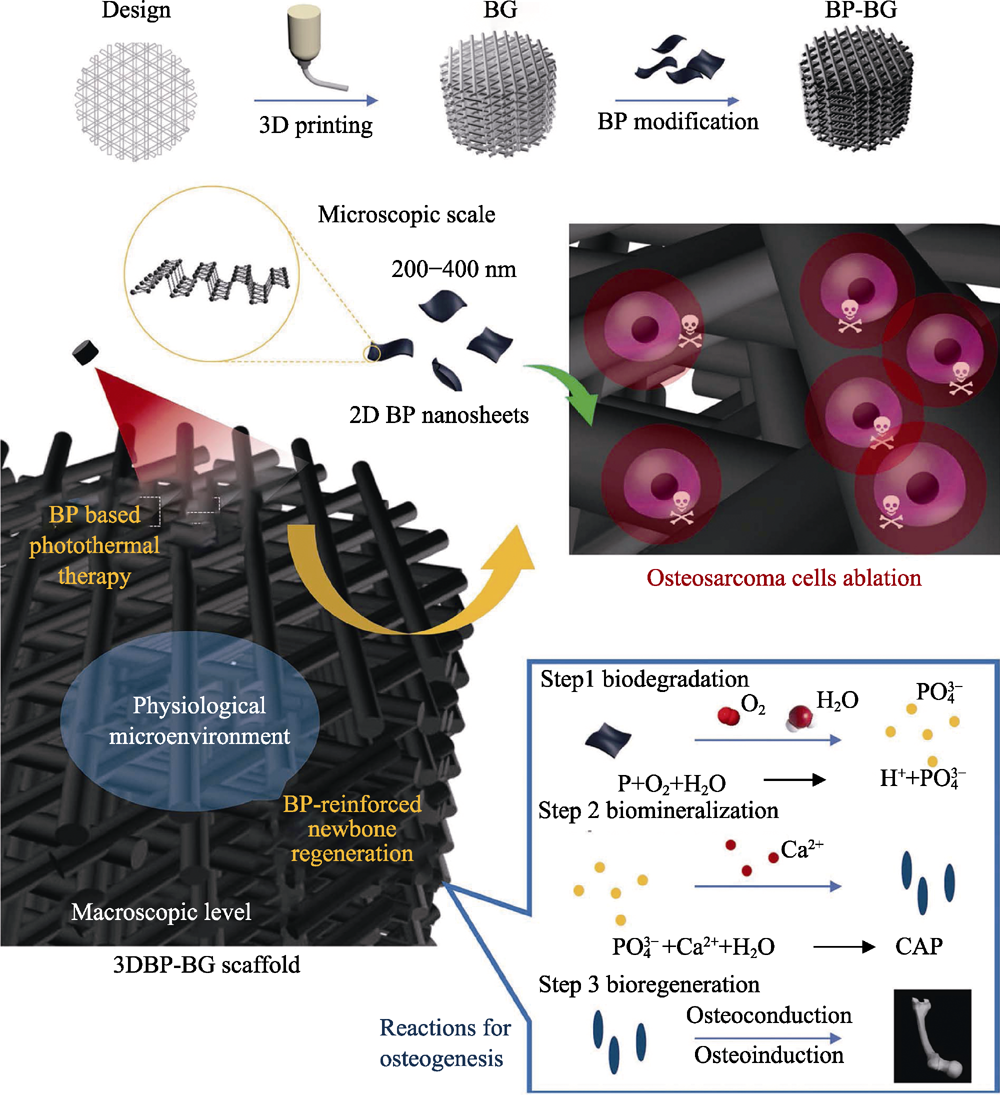
Fig. 7 Schematic illustration of fabrication process of BP-BG scaffold and stepwise therapeutic strategy for the elimination of osteosarcoma followed by osteogenesis by BP-BG[16]
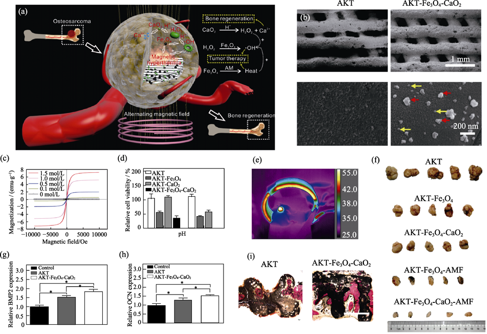
Fig. 8 (a) Schematic diagram of AKT-Fe3O4-CaO2 scaffold functioning to obtain efficient tumor ablation and enhanced bone regeneration, (b) SEM images of AKT and AKT-Fe3O4-CaO2 scaffold (red arrows: Fe3O4 nanoparticles; yellow arrows: CaO2 nanoparticles), (c) magnetization curves of AKT-Fe3O4-CaO2 scaffolds soaked in Fe3O4 suspensions with various concentrations, (d) in vitro therapeutic effect of AKT-Fe3O4-CaO2 scaffold, (e) infrared images of nude mice in alternating magnetic fields, (f) in vivo therapeutic effect of AKT-Fe3O4-CaO2 scaffold, (g, h) expression of osteogenic genes of BMSCs, and (i) regeneration of cranium defects implanted with AKT and AKT-Fe3O4-CaO2 scaffolds[7] (1 emu?g-1= 1×10³ A?m-1?g-1, 1 Oe=80 A?m-1)
| [1] |
GEORGAKILAS V, TIWARI J N, KEMP K C, et al. Noncovalent functionalization of graphene and graphene oxide for energy materials, biosensing, catalytic, and ciomedical applications. Chemical Reviews, 2016,116(9):5464-5519.
DOI URL PMID |
| [2] |
HABRAKEN W, HABIBOVIC P, EPPLE M, et al. Calcium phosphates in biomedical applications: materials for the future? Materials Today, 2016,19(2):69-87.
DOI URL |
| [3] |
LIN K L, WU C T, CHANG J. Advances in synthesis of calcium phosphate crystals with controlled size and shape. Acta Biomaterialia, 2014,10(10):4071-4102.
DOI URL |
| [4] |
WANG X L, REN Z M, CHANG J. Synthesis and orientation of Fe-doped hydroxyapatite in high magnetic field. Journal of Inorganic Materials, 2018,33(1):75-80.
DOI URL |
| [5] |
ZHANG B, YANG C A, SHI P. Synthesis of graphene/ hydroxyapatite composite bioceramics via plasma Activated sintering, Journal of Inorganic Materials, 2018,33(12):1355-1359.
DOI URL |
| [6] |
WU C T, CHANG J. Silicate bioceramics for bone tissue regeneration. Journal of Inorganic Materials, 2013,28(1):29-39.
DOI URL |
| [7] | DONG S, CHEN Y, YU L, et al. Magnetic hyperthermia- synergistic H2O2 self-sufficient catalytic suppression of osteosarcoma with enhanced bone-regeneration bioactivity by 3D-printing composite scaffolds. Advanced Functional Materials, 2019, 30(4): 1907071-1-15. |
| [8] |
SHI Z Y, LI Q, TANG S C, et al. Surface modification on property of mesoporous calcium magnesium silicate/polyetheretherketone composites. Journal of Inorganic Materials, 2018,33(1):67-74.
DOI URL |
| [9] |
BAI F, WANG Z, LU J, et al. The correlation between the internal structure and vascularization of controllable porous bioceramic aterials in vivo: a quantitative study. Tissue Engineering Part A, 2010,16(12):3791-3803.
DOI URL PMID |
| [10] |
LIN K L, CHANG J, ZENG Y, et al. Preparation of macroporous calcium silicate ceramics. Materials Letters, 2004,58(15):2109-2113.
DOI URL |
| [11] |
BAINO F, FIUME E, BARBERI J, et al. Processing methods for making porous bioactive glass-based scaffolds-a state-of-the-art review. International Journal of Applied Ceramic Technology, 2019,16(5):1762-1796.
DOI URL |
| [12] |
XIN C, QI X, ZHU M, et al. Hydroxyapatite whisker-reinforced composite scaffolds through 3D printing for bone repair. Journal of Inorganic Materials, 2017,32(8):837-844.
DOI URL |
| [13] |
MA H, FENG C, CHANG J, et al. 3D-printed bioceramic scaffolds: from bone tissue engineering to tumor therapy. Acta Biomaterialia, 2018,79:37-59.
DOI URL PMID |
| [14] |
VON ERLACH T C, BERTAZZO S, WOZNIAK M A, et al. Cell-geometry-dependent changes in plasma membrane order direct stem cell signalling and fate. Nature Materials, 2018,17(3):237-242.
DOI URL PMID |
| [15] | ROGOWSKA-TYLMAN J, LOCS J, SALMA I, et al. In vivo and in vitro study of a novel nanohydroxyapatite sonocoated scaffolds for enhanced bone regeneration. Materials Science & Engineering: C, 2019,99:669-684. |
| [16] | YANG B W, YIN J H, CHEN Y, et al. 2D-black-phosphorus- reinforced 3D-printed scaffolds: a stepwise countermeasure for osteosarcoma. Advanced Materials, 2018, 30(10): 1705611-1-12. |
| [17] | ZHANG Y L, ZHAI D, XU M C, et al. 3D-printed bioceramic scaffolds with antibacterial and osteogenic activity. Biofabrication, 2017, 9(2): 025037-1-12. |
| [18] |
ZHANG X, LI H, LIU J, et al. Amorphous carbon modification on implant surface: a general strategy to enhance osteogenic differentiation for diverse biomaterials via FAK/ERK1/2 signaling pathways. Journal of Materials Chemistry B, 2019,7(15):2518-2533.
DOI URL PMID |
| [19] |
TOURI M, MOZTARZADEH F, OSMAN N A A, et al. 3D- printed biphasic calcium phosphate scaffolds coated with an oxygen generating system for enhancing engineered tissue survival. Materials Science and Engineering: C, 2018,84:236-242.
DOI URL |
| [20] |
WANG C, LIN K, CHANG J, et al. Osteogenesis and angiogenesis induced by porous beta-CaSiO3/PDLGA composite scaffold via activation of AMPK/ERK1/2 and PI3K/Akt pathways. Biomaterials, 2013,34(1):64-77.
DOI URL |
| [21] |
LIN K, XIA L, GAN J, et al. Tailoring the nanostructured surfaces of hydroxyapatite bioceramics to promote protein adsorption, osteoblast growth, and osteogenic differentiation. ACS Applied Materials & Interfaces, 2013,5(16):8008-8017.
URL PMID |
| [22] |
ZHAO C, XIA L, ZHAI D, et al. Designing ordered micropatterned hydroxyapatite bioceramics to promote the growth and osteogenic differentiation of bone marrow stromal cells. Journal of Materials Chemistry B, 2015,3(6):968-976.
DOI URL PMID |
| [23] |
MAO L, LIU J, ZHAO J, et al. Effect of micro-nano-hybrid structured hydroxyapatite bioceramics on osteogenic and cementogenic differentiation of human periodontal ligament stem cell via Wnt signaling pathway. International Journal of Nanomedicine, 2015,10:7031-7044.
DOI URL PMID |
| [24] |
XIA L, LIN K, JIANG X, et al. Effect of nano-structured bioceramic surface on osteogenic differentiation of adipose derived stem cells. Biomaterials, 2014,35(30):8514-8527.
DOI URL |
| [25] |
WANG X, ZHOU Y, XIA L, et al. Fabrication of nano-structured calcium silicate coatings with enhanced stability, bioactivity and osteogenic and angiogenic activity. Colloids and Surfaces B-Biointerfaces, 2015,126:358-366.
DOI URL |
| [26] |
XIA L, XIE Y, FANG B, et al. In situ modulation of crystallinity and nano-structures to enhance the stability and osseointegration of hydroxyapatite coatings on Ti-6Al-4V implants. Chemical Engineering Journal, 2018,347:711-720.
DOI URL |
| [27] |
XIA L, ZHANG N, WANG X, et al. The synergetic effect of nano-structures and silicon-substitution on the properties of hydroxyapatite scaffolds for bone regeneration. Journal of Materials Chemistry B, 2016,4(19):3313-3323.
DOI URL PMID |
| [28] |
DENG C J, LIN R C, ZHANG M, et al. Micro/nanometer- structured scaffolds for regeneration of both cartilage and subchondral bone. Advanced Functional Materials, 2019,29(4):1806068-15.
DOI URL |
| [29] |
YANG C, WANG X Y, MA B, et al. 3D-printed bioactive Ca3SiO5 bone cement scaffolds with nano surface structure for bone regeneration. ACS Applied Materials & Interfaces, 2017,9(7):5757-5767.
DOI URL PMID |
| [30] | WANG X C, LI T, MA H S, et al. A 3D-printed scaffold with MoS2 nanosheets for tumor therapy and tissue regeneration. NPG Asia Materials, 2017, 9: e376-1-14. |
| [31] |
XIA L, LIN K, JIANG X, et al. Enhanced osteogenesis through nano-structured surface design of macroporous hydroxyapatite bioceramic scaffolds via activation of ERK and p38 MAPK signaling pathways. Journal of Materials Chemistry B, 2013,1(40):5403-5416.
DOI URL |
| [32] |
DONG S, ZHANG Y-N, WANG J, et al. A novel multifunctional carbon aerogel coated platform for osteosarcoma therapy and enhanced bone regeneration. Journal of Materials Chemistry B, 2020,8(3):368-379.
DOI URL PMID |
| [33] |
WANG C, LIN K, CHANG J, et al. The stimulation of osteogenic differentiation of mesenchymal stem cells and vascular endothelial growth factor secretion of endothelial cells by beta-CaSiO3/beta- Ca3(PO4)(2) scaffolds. Journal of Biomedical Materials Research Part A, 2014,102(7):2096-2104.
DOI URL |
| [34] |
WITTE F, KAESE V, HAFERKAMP H, et al. In vivo corrosion of four magnesium alloys and the associated bone response. Biomaterials, 2005,26(17):3557-3563.
DOI URL |
| [35] |
YOSHIZAWA S, BROWN A, BARCHOWSKY A, et al. Magnesium ion stimulation of bone marrow stromal cells enhances osteogenic activity, simulating the effect of magnesium alloy degradation. Acta Biomaterialia, 2014,10(6):2834-2842.
DOI URL |
| [36] | XIA L, YIN Z, MAO L, et al. Akermanite bioceramics promote osteogenesis,angiogenesis and suppress osteoclastogenesis for osteoporotic bone regeneration. Scientific Reports, 2016, 6: 22005- 1-17. |
| [37] |
AMIN N, CLARK C C T, TAGHIZADEH M, et al. Zinc supplements and bone health: the role of the RANKL-RANK axis as a therapeutic target. Journal of Trace Elements in Medicine and Biology, 2019, DOI: 10.1016/j.jtemb.2019.126417.
DOI URL PMID |
| [38] |
QIAO Y Q, ZHANG W J, TIAN P, et al. Stimulation of bone growth following zinc incorporation into biomaterials. Biomaterials, 2014,35(25):6882-6897.
DOI URL |
| [39] |
LIN K L, XIA L G, LI H Y, et al. Enhanced osteoporotic bone regeneration by strontium-substituted calcium silicate bioactive ceramics. Biomaterials, 2013,34(38):10028-10042.
DOI URL |
| [40] |
LIU W, WANG T, YANG C, et al. Alkaline biodegradable implants for osteoporotic bone defects-importance of microenvironment pH. Osteoporosis International, 2016,27(1):93-104.
DOI URL PMID |
| [41] |
GUO X, WEI S, LU M, et al. Dose-dependent effects of strontium ranelate on ovariectomy rat bone marrow mesenchymal stem cells and human umbilical vein endothelial cells. International Journal of Biological Sciences, 2016,12(12):1511-1522.
DOI URL PMID |
| [42] | GUO X, WEI S, LU M, et al. RNA-Seq investigation and in vivo study the effect of strontium ranelate on ovariectomized rat via the involvement of ROCK1. Artificial Cells Nanomedicine and Biotechnology, 2018,46:S629-S641. |
| [43] |
LIN K, WANG X, ZHANG N, et al. Strontium (Sr) strengthens the silicon (Si) upon osteoblast proliferation, osteogenic differentiation and angiogenic factor expression. Journal of Materials Chemistry B, 2016,4(21):3632-3638.
DOI URL PMID |
| [44] | WANG C Y, CHEN B, WANG W, et al. Strontium released bi-lineage scaffolds with immunomodulatory properties induce a pro-regenerative environment for osteochondral regeneration. Materials Science & Engineering: C, 2019, 103: 109833-1-12. |
| [45] |
ZHANG X, LI H, LIN C, et al. Synergetic topography and chemistry cues guiding osteogenic differentiation in bone marrow stromal cells through ERK1/2 and p38 MAPK signaling pathway. Biomaterials Science, 2018,6(2):418-430.
DOI URL PMID |
| [46] |
FIELDING G, BOSE S. SiO2 and ZnO dopants in three- dimensionally printed tricalcium phosphate bone tissue engineering scaffolds enhance osteogenesis and angiogenesis in vivo. Acta Biomaterialia, 2013,9(11):9137-9148.
DOI URL PMID |
| [47] |
WU C, ZHAI D, MA H, et al. Stimulation of osteogenic and angiogenic ability of cells on polymers by pulsed laser deposition of uniform akermanite-glass nanolayer. Acta Biomaterialia, 2014,10(7):3295-3306.
DOI URL |
| [48] |
KONG N, LIN K, LI H, et al. Synergy effects of copper and silicon ions on stimulation of vascularization by copper-doped calcium silicate. Journal of Materials Chemistry B, 2014,2(8):1100-1110.
DOI URL |
| [49] |
SHI M C, CHEN Z T, FARNAGHI S, et al. Copper-doped mesoporous silica nanospheres, a promising immunomodulatory agent for inducing osteogenesis. Acta Biomaterialia, 2016,30:334-344.
DOI URL PMID |
| [50] |
LI J Y, ZHAI D, LV F, et al. Preparation of copper-containing bioactive glass/eggshell membrane nanocomposites for improving angiogenesis, antibacterial activity and wound healing. Acta Biomaterialia, 2016,36:254-266.
DOI URL PMID |
| [51] |
LI H, LI J Y, JIANG J, et al. An osteogenesis/angiogenesis- stimulation artificial ligament for anterior cruciate ligament reconstruction. Acta Biomaterialia, 2017,54:399-410.
DOI URL PMID |
| [52] |
ZHOU Y H, HAN S W, XIAO L, et al. Accelerated host angiogenesis and immune responses by ion release from mesoporous bioactive glass. Journal of Materials Chemistry B, 2018,6(20):3274-3284.
DOI URL PMID |
| [53] |
LIN R C, DENG C J, LI X X, et al. Copper-incorporated bioactive glass-ceramics inducing anti-inflammatory phenotype and regeneration of cartilage/bone interface. Theranostics, 2019,9(21):6300-6313.
DOI URL PMID |
| [54] |
DANG W T, WANG X Y, LI J Y, et al. 3D printing of Mo- containing scaffolds with activated anabolic responses and bi-lineage bioactivities. Theranostics, 2018,8(16):4372-4392.
DOI URL PMID |
| [55] |
LIU Y Q, LI T, MA H S, et al. 3D-printed scaffolds with bioactive elements-induced photothermal effect for bone tumor therapy. Acta Biomaterialia, 2018,73:531-546.
DOI URL PMID |
| [56] |
MEININGER S, MANDAL S, KUMAR A, et al. Strength reliability and in vitro degradation of three-dimensional powder printed strontium-substituted magnesium phosphate scaffolds. Acta Biomaterialia, 2016,31:401-411.
DOI URL PMID |
| [57] |
MEININGER S, MOSEKE C, SPATZ K, et al. Effect of strontium substitution on the material properties and osteogenic potential of 3D powder printed magnesium phosphate scaffolds. Materials Science and Engineering: C, 2019,98:1145-1158.
DOI URL |
| [58] |
XIE J, SHAO H, HE D, et al. Ultrahigh strength of three- dimensional printed diluted magnesium doping wollastonite porous scaffolds. MRS Communications, 2015,5(4):631-639.
DOI URL |
| [59] |
KE X, ZHANG L, YANG X, et al. Low-melt bioactive glass- reinforced 3D printing akermanite porous cages with highly improved mechanical properties for lumbar spinal fusion. J Tissue Eng. Regen. Med., 2018,12(5):1149-1162.
DOI URL PMID |
| [60] |
FU S Y, YU B, DING H F, et al, Zirconia incorporation in 3D printed beta-Ca2SiO4 scaffolds on their physicochemical and biological property. Journal of Inorganic Materials, 2019,34(4):444-454.
DOI URL |
| [61] |
ZHANG F, CHANG J, LU J, et al. Bioinspired structure of bioceramics for bone regeneration in load-bearing sites. Acta Biomaterialia, 2007,3(6):896-904.
DOI URL |
| [62] | FENG C, ZHANG W, DENG C, et al. 3D Printing of lotus root-like biomimetic materials for cell delivery and tissue regeneration. Advanced Science, 2017, 4(12): 1700401-1-9. |
| [63] |
ALMELA T, BROOK I M, KHOSHROO K, et al. Simulation of cortico-cancellous bone structure by 3D printing of bilayer calcium phosphate-based scaffolds. Bioprinting, 2017,6:1-7.
DOI URL |
| [64] |
JUNG-BIN L, WOO-YOUL M, YOUNG-HAG K, et al. Porous calcium phosphate ceramic scaffolds with tailored pore orientations and mechanical properties using lithography-based ceramic 3D printing technique. Materials, 11(9):1711-1718.
DOI URL |
| [65] |
MATHARU G S, DANIEL J, ZIAEE H, et al. Failure of a novel ceramic-on-ceramic hip resurfacing prosthesis. Journal of Arthroplasty, 2015,30(3):416-418.
DOI URL PMID |
| [66] |
MANNY P, JAVAD P, SHARKEY P F, et al. Causes of failure of ceramic-on-ceramic and metal-on-metal hip arthroplasties. Clinical Orthopaedics & Related Research, 2012,470(2):382-387.
DOI URL PMID |
| [67] |
ZHAO L, WU C T, LIN K L, et al. The effect of poly(lactic-co- glycolic acid)(PLGA) coating on the mechanical, biodegradable, bioactive properties and drug release of porous calcium silicate scaffolds. Bio-medical Materials and Engineering, 2012,22(5):289-300.
DOI URL |
| [68] |
LI CHEN, AI FANRONG, MIAO XINXIN, et al. “The return of ceramic implants”: rose stem inspired dual layered modification of ceramic scaffolds with improved mechanical and anti-infective properties. Materials Science and Engineering: C, 2018,93:873-879.
DOI URL |
| [69] | KIM B S, YANG S S, PARK H, et al. Improvement of mechanical strength and osteogenic potential of calcium sulfate-based hydroxyapatite 3-dimensional printed scaffolds by epsilon- polycarbonate coating. Journal of Biomaterials Science-Polymer Edition, 2017,28(13):1256-1270. |
| [70] |
DíAZ-RODRíGUEZ P, GONZáLEZ P, SERRA J, et al. Key parameters in blood-surface interactions of 3D bioinspired ceramic materials. Materials Science and Engineering: C, 2014,41:232-239.
DOI URL |
| [71] |
LIENEMANN P S, LUTOLF M P, MARTIN E. Biomimetic hydrogels for controlled biomolecule delivery to augment bone regeneration. Advanced Drug Delivery Reviews, 2012,64(12):1078-1089.
DOI URL PMID |
| [72] |
LI M, CHENG Y, ZHENG Y F, et al. Surface characteristics and corrosion behaviour of WE43 magnesium alloy coated by SiC film. Applied Surface Science. 2012,258(7):3074-3081.
DOI URL |
| [73] |
YANG Y, LAI Y, ZHANG Q, et al. A novel electrochemical strategy for improving blood compatibility of titanium-based biomaterials. Colloids & Surfaces B Biointerfaces, 2010,79(1):309-313.
DOI URL PMID |
| [74] |
ZHAO L, LIN K L, ZHANG M L, et al. The influences of poly(lactic-co-glycolic acid) (PLGA) coating on the biodegradability, bioactivity, and biocompatibility of calcium silicate bioceramics. Journal of Materials Science, 2011,46(14):4986-4993.
DOI URL |
| [75] |
WEI T, DANG-SHENG X. Bioinspired superhydrophobic progress and recent advances of its functional application. Journal of Inorganic Materials, 2019,34(11):1133-1144.
DOI URL |
| [76] |
ARIMA Y, IWATA H, Effect of wettability and surface functional groups on protein adsorption and cell adhesion using well-defined mixed self-assembled monolayers. Biomaterials, 2007,28(20):3074-3082.
DOI URL |
| [77] |
TENG Y Q, ZHANG Y Q, HENG L P, et al. Conductive polymer porous film with tunable wettability and adhesion. Materials, 2015,8(4):1817-1830.
DOI URL PMID |
| [78] |
CHEN Z, ZHANG D, PENG E, et al. 3D-printed ceramic structures with in situ grown whiskers for effective oil/water separation. Chemical Engineering Journal, 2019,373:1223-1232.
DOI URL |
| [79] |
SONG Y, LIN K, HE S, et al. Nano-biphasic calcium phosphate/polyvinyl alcohol composites with enhanced bioactivity for bone repair via low-temperature three-dimensional printing and loading with platelet-rich fibrin. International Journal of Nanomedicine, 2018,13:505-523.
DOI URL PMID |
| [80] |
TROMBETTA R, INZANA J A, SCHWARZ E M, et al. 3D printing of calcium phosphate ceramics for bone tissue engineering and drug delivery. Annals of Biomedical Engineering, 2017,45(1):1-22.
DOI URL PMID |
| [81] |
BOSE S, VAHABZADEH S, BANDYOPADHYAY A. Bone tissue engineering using 3D printing. Materials Today, 2013,16(12):496-504.
DOI URL |
| [82] |
ZHANG Y L, ZHAI D, XU M C, et al. 3D-printed bioceramic scaffolds with a Fe3O4/graphene oxide nanocomposite interface for hyperthermia therapy of bone tumor cells. Journal of Materials Chemistry B, 2016,4(17):2874-2886.
DOI URL PMID |
| [83] |
TORRES P M C, VIEIRA S I, CERQUEIRA A R, et al. Effects of Mn-doping on the structure and biological properties of β-tricalcium phosphate. Journal of Inorganic Biochemistry, 2014,136:57-66.
DOI URL |
| [84] |
MIN Z, SHICHANG Z, CHEN X, et al. 3D-printed dimethyloxallyl glycine delivery scaffolds to improve angiogenesis and osteogenesis. Biomater. Sci., 2015,3(8):1236-1244.
DOI URL PMID |
| [85] | DIAZ-RODRIGUEZ P, SANCHEZ M, LANDIN M. Drug-loaded biomimetic ceramics for tissue engineering. Pharmaceutics, 2018, 10(4): 272-1-20. |
| [86] |
MEIßNER R, BERTOL L, REHMAN M A U, et al. Bioprinted 3D calcium phosphate scaffolds with gentamicin releasing capability. Ceramics International, 2019,45(6):7090-7094.
DOI URL |
| [87] |
LI T, ZHAI D, MA B, et al. 3D printing of hot dog-like biomaterials with hierarchical architecture and distinct bioactivity. Advanced Science, 2019,6(19):1901146-8.
DOI URL PMID |
| [88] |
SONG J-J, CHEN B, LIN K-L. Core-shell structured hydroxyapatite/mesoporous silica nanoparticle: preparation and application in drug delivery. Journal of Inorganic Materials, 2018,33(6):623-628.
DOI URL |
| [89] |
CHEN G, ROY I, YANG C, et al. Nanochemistry and nanomedicine for nanoparticle-based diagnostics and therapy. Chemical Reviews, 2016,116(5):2826-2885.
DOI URL PMID |
| [90] | ZHEN F, YAN C, CUI C, et al. Near infrared fluorescent peptide nanoparticles for enhancing esophageal cancer therapeutic efficacy. Nature Communications, 2018, 9(1): 2605-1-11. |
| [91] |
WANG S, CHEN Y, LI X, et al. Injectable 2D MoS2-integrated drug delivering implant for highly efficient NIR-triggered synergistic tumor hyperthermia. Advanced Materials, 2015,27(44):7117-7122.
DOI URL PMID |
| [92] | CHANDRAWATI D R, CHANG D J Y H, REINATORRES D E, et al. Localized and controlled delivery of nitric oxide to the conventional outflow pathway via enzyme biocatalysis: toward therapy for glaucoma. Advanced Materials, 2017, 29(16): 1604932-1-7. |
| [93] |
BADGWELL B, BLUM M, ESTRELLA J, et al. Personalised therapy for localised gastric and gastro-oesophageal adenocarcinoma. Lancet Oncology, 2016,17(12):1628-1629.
DOI URL PMID |
| [94] |
YANG B W, GU Z, CHEN Y. Nanomedicine-augmented cancer-localized treatment by 3D theranostic implants. Journal of Biomedical Nanotechnology, 2017,13(8):871-890.
DOI URL |
| [95] |
MA H, JIANG C, ZHAI D, et al. A bifunctional biomaterial with photothermal effect for tumor therapy and bone regeneration. Advanced Functional Materials, 2016,26(8):1197-1208.
DOI URL |
| [96] |
WU P, GRAINGER D W. Drug/device combinations for local drug therapies and infection prophylaxis. Biomaterials, 2006,27(11):2450-2467.
DOI URL |
| [97] |
ARIJIT KUMAR C, RUCHIRA C, TARAKDAS B. Mechanism of antibacterial activity of copper nanoparticles. Nanotechnology, 2014, 25(13): 135101-1-13.
DOI URL PMID |
| [98] |
SCHRAND A M, RAHMAN M F, HUSSAIN S M, et al. Metal-based nanoparticles and their toxicity assessment. Wiley Interdisciplinary Reviews: Nanomedicine and Nanobiotechnology, 2010,2(5):544-568.
DOI URL PMID |
| [99] |
LIAO F, MA J Q, GE H G. Preparation, characterization and antimicrobial activity of core-satellite Ag/PDA@SiO2@CoFe2O4 magnetic composites. Journal of Inorganic Materials, 2017,32(5):523-528.
DOI URL |
| [100] |
LIU Y L, WANG R L, LI N, et al. Preparation of zinc oxide mesocrystal filler and the properties of dental composite resins. Journal of Inorganic Materials, 2019,34(10):1077-1084.
DOI URL |
| [101] |
WANG Q, TANG P F, GE X, et al. Experimental and simulation studies of strontium/zinc-codoped hydroxyapatite porous scaffolds with excellent osteoinductivity and antibacterial activity. Applied Surface Science, 2018,462:118-126.
DOI URL |
| [102] |
VALAPPIL S P, COOMBES M, WRIGHT L, et al. Role of gallium and silver from phosphate-based glasses on in vitro dual species oral biofilm models of porphyromonas gingivalis and Streptococcus gordonii. Acta Biomaterialia, 2012,8(5):1957-1965.
DOI URL |
| [103] |
GOH Y F, ALSHEMARY A Z, AKRAM M, et al. In-vitro characterization of antibacterial bioactive glass containing ceria. Ceramics International, 2014,40(1):729-737.
DOI URL |
| [104] |
CHATZISTAVROU X, FENNO J C, FAULK D, et al. Fabrication and characterization of bioactive and antibacterial composites for dental applications. Acta Biomaterialia, 2014,10(8):3723-3732.
DOI URL |
| [105] | BRAUER D S, KARPUKHINA N, KEDIA G, et al. Bactericidal strontium-releasing injectable bone cements based on bioactive glasses. Journal of the Royal Society Interface, 2013, 10(78): 20120647-1-8. |
| [106] |
WU Y, XIA L, ZHOU Y, et al. Evaluation of osteogenesis and angiogenesis of icariin loaded on micro/nano hybrid structured hydroxyapatite granules as a local drug delivery system for femoral defect repair. Journal of Materials Chemistry B, 2015,3(24):4871-4883.
DOI URL PMID |
| [107] |
ZHOU Y, WU Y, MA W, et al. The effect of quercetin delivery system on osteogenesis and angiogenesis under osteoporotic conditions. Journal of Materials Chemistry B, 2017,5(3):612-625.
DOI URL PMID |
| [108] | ZHANG F M, CHANG J, LIN K L, et al. Preparation, mechanical properties and in vitro degradability of wollastonite/tricalcium phosphate macroporous scaffolds from nanocomposite powders. Journal of Materials Science-Materials in Medicine, 2008,19(1):167-173. |
| [109] |
ZHANG F, LIN K, CHANG J, et al. Spark plasma sintering of macroporous calcium phosphate scaffolds from nanocrystalline powders. Journal of The European Ceramic Society, 2008,28(3):539-545.
DOI URL |
| [1] | ZHU Wenjie, TANG Lu, LU Jichang, LIU Jiangping, LUO Yongming. Research Progress on Catalytic Oxidation of Volatile Organic Compounds by Perovskite Oxides [J]. Journal of Inorganic Materials, 2025, 40(7): 735-746. |
| [2] | SUN Jing, LI Xiang, MAO Xiaojian, ZHANG Jian, WANG Shiwei. Effect of Lauric Acid Modifier on the Hydrolysis Resistance of Aluminum Nitride Powders [J]. Journal of Inorganic Materials, 2025, 40(7): 826-832. |
| [3] | HU Zhichao, YANG Hongyu, YANG Hongcheng, SUN Chengli, YANG Jun, LI Enzhu. Usage of the P-V-L Bond Theory in Regulating Properties of Microwave Dielectric Ceramics [J]. Journal of Inorganic Materials, 2025, 40(6): 609-626. |
| [4] | WU Qiong, SHEN Binglin, ZHANG Maohua, YAO Fangzhou, XING Zhipeng, WANG Ke. Research Progress on Lead-based Textured Piezoelectric Ceramics [J]. Journal of Inorganic Materials, 2025, 40(6): 563-574. |
| [5] | ZHANG Bihui, LIU Xiaoqiang, CHEN Xiangming. Recent Progress of Hybrid Improper Ferroelectrics with Ruddlesden-Popper Structure [J]. Journal of Inorganic Materials, 2025, 40(6): 587-608. |
| [6] | WU Jie, YANG Shuai, WANG Mingwen, LI Jinglei, LI Chunchun, LI Fei. Textured PT-based Piezoelectric Ceramics: Development, Status and Challenge [J]. Journal of Inorganic Materials, 2025, 40(6): 575-586. |
| [7] | JIANG Kun, LI Letian, ZHENG Mupeng, HU Yongming, PAN Qinxue, WU Chaofeng, WANG Ke. Research Progress on Low-temperature Sintering of PZT Ceramics [J]. Journal of Inorganic Materials, 2025, 40(6): 627-638. |
| [8] | CHEN Xi, YUAN Yuan, TAN Yeqiang, LIU Changsheng. Strategic Study on the Development of Inorganic Non-metallic Biomaterials [J]. Journal of Inorganic Materials, 2025, 40(5): 449-456. |
| [9] | TIAN Ruizhi, LAN Zhengyi, YIN Jie, HAO Nanjing, CHEN Hangrong, MA Ming. Microfluidic Technology Based Synthesis of Inorganic Nano-biomaterials: Principles and Progress [J]. Journal of Inorganic Materials, 2025, 40(4): 337-347. |
| [10] | ZHANG Jiguo, WU Tian, ZHAO Xu, YANG Fan, XIA Tian, SUN Shien. Improvement of Cycling Stability of Cathode Materials and Industrialization Process for Sodium-ion Batteries [J]. Journal of Inorganic Materials, 2025, 40(4): 348-362. |
| [11] | YIN Jie, GENG Jiayi, WANG Kanglong, CHEN Zhongming, LIU Xuejian, HUANG Zhengren. Recent Advances in 3D Printing and Densification of SiC Ceramics [J]. Journal of Inorganic Materials, 2025, 40(3): 245-255. |
| [12] | CHEN Guangchang, DUAN Xiaoming, ZHU Jinrong, GONG Qing, CAI Delong, LI Yuhang, YANG Donglei, CHEN Biao, LI Xinmin, DENG Xudong, YU Jin, LIU Boya, HE Peigang, JIA Dechang, ZHOU Yu. Advanced Ceramic Materials in Helicopter Special Structures: Research Progress and Application Prospect [J]. Journal of Inorganic Materials, 2025, 40(3): 225-244. |
| [13] | FAN Xiaobo, ZU Mei, YANG Xiangfei, SONG Ce, CHEN Chen, WANG Zi, LUO Wenhua, CHENG Haifeng. Research Progress on Proton-regulated Electrochemical Ionic Synapses [J]. Journal of Inorganic Materials, 2025, 40(3): 256-270. |
| [14] | HAIREGU Tuxun, GUO Le, DING Jiayi, ZHOU Jiaqi, ZHANG Xueliang, NUERNISHA Alifu. Research Progress of Optical Bioimaging Technology Assisted by Upconversion Fluorescence Probes in Tumor Imaging [J]. Journal of Inorganic Materials, 2025, 40(2): 145-158. |
| [15] | SUN Shujuan, ZHENG Nannan, PAN Haokun, MA Meng, CHEN Jun, HUANG Xiubing. Research Progress on Preparation Methods of Single-atom Catalysts [J]. Journal of Inorganic Materials, 2025, 40(2): 113-127. |
| Viewed | ||||||
|
Full text |
|
|||||
|
Abstract |
|
|||||